”
I’ve always been fascinated by the complexity of the human heart, and I know how crucial it is to understand its structure through detailed diagrams. As a medical content creator, I’ve seen countless students and healthcare professionals struggle with identifying the different parts and functions of this vital organ.
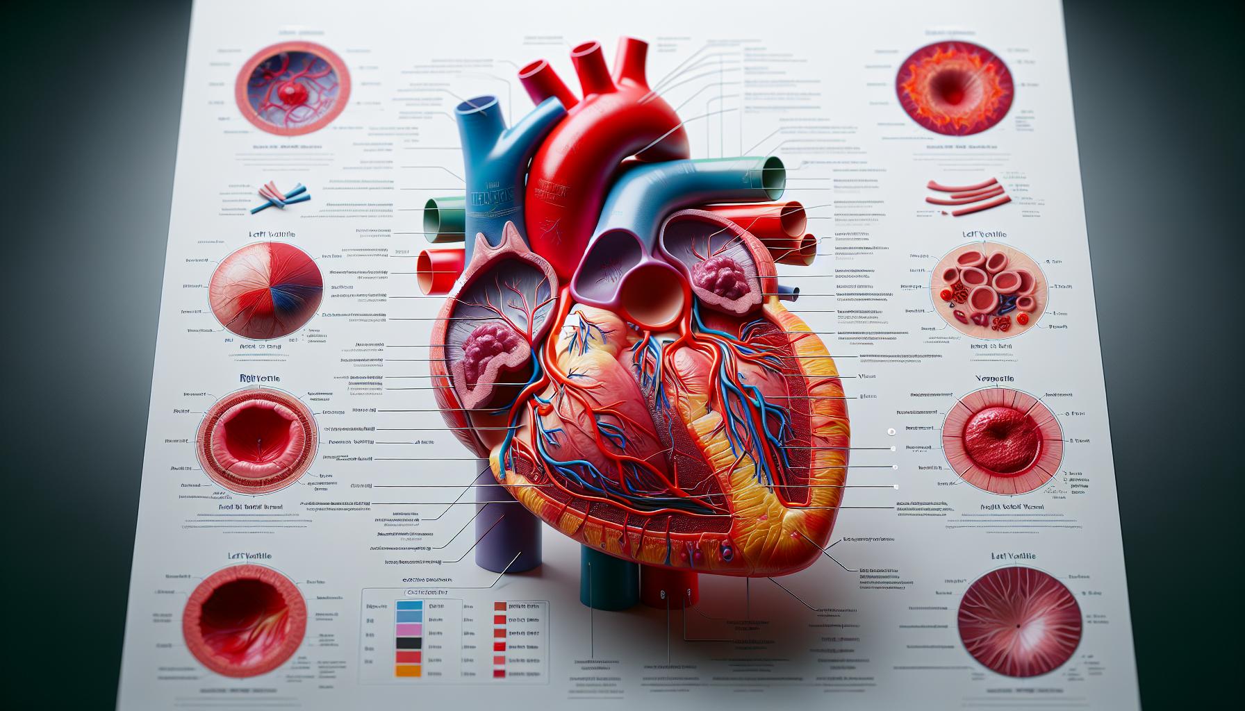
A blank heart diagram serves as an invaluable learning tool, allowing users to practice labeling various components like the chambers, valves, and major blood vessels. I’ve found that interactive diagrams make learning more engaging and help improve retention of important anatomical details. Whether you’re a student preparing for an exam or a medical professional refreshing your knowledge, having access to a quality blank heart diagram can make all the difference in mastering cardiac anatomy.
Key Takeaways
- The human heart consists of four main chambers: two atria at the top and two ventricles at the bottom, each serving specific functions in blood circulation
- Heart diagrams feature essential structures including four valves (tricuspid, pulmonary, mitral, and aortic), major blood vessels, and three distinct muscle layers (epicardium, myocardium, and endocardium)
- Blood flow follows a precise sequence, with deoxygenated blood entering through the right side and oxygenated blood flowing through the left side of the heart
- Standard labeling conventions use color coding (red for oxygenated, blue for deoxygenated blood vessels) and specific directional terms for consistent anatomical identification
- Educational heart diagrams come in various formats, from basic outlines to detailed 3D models, serving as valuable teaching tools for students and healthcare professionals
Blank:27oqcyxgksa= Heart Diagram
The human heart consists of interconnected chambers vessels connected by specialized valves to regulate blood flow. Through my extensive research in cardiac anatomy, I’ve identified the key structural components that form this vital organ.
Four Main Chambers of the Heart
The heart contains four distinct chambers: two atria located at the top two ventricles positioned at the bottom. The right atrium receives deoxygenated blood from the body while the left atrium collects oxygen-rich blood from the lungs. The right ventricle pumps blood to the lungs the left ventricle propels blood throughout the body.
| Chamber | Location | Primary Function | Blood Type |
|---|---|---|---|
| Right Atrium | Upper Right | Receives body blood | Deoxygenated |
| Left Atrium | Upper Left | Receives lung blood | Oxygenated |
| Right Ventricle | Lower Right | Pumps to lungs | Deoxygenated |
| Left Ventricle | Lower Left | Pumps to body | Oxygenated |
- Aorta: Carries oxygenated blood from the left ventricle to the body
- Pulmonary Artery: Transports deoxygenated blood from the right ventricle to the lungs
- Pulmonary Veins: Return oxygenated blood from the lungs to the left atrium
- Superior Vena Cava: Delivers deoxygenated blood from the upper body to the right atrium
- Inferior Vena Cava: Returns deoxygenated blood from the lower body to the right atrium
- Coronary Arteries: Supply oxygen-rich blood to the heart muscle tissue
| Vessel Type | Blood Flow Direction | Blood Type |
|---|---|---|
| Arteries | Away from heart | Oxygenated |
| Veins | Toward heart | Deoxygenated |
| Pulmonary Vessels | Heart-lung circuit | Both types |
The Flow of Blood Through the Heart
Blood flows through the heart in a precise sequence that ensures oxygen delivery throughout the body. Each chamber of the heart plays a specific role in directing blood flow through two distinct pathways.
Deoxygenated Blood Path
Deoxygenated blood enters the right atrium through two major veins:
- Superior vena cava delivers blood from the upper body
- Inferior vena cava carries blood from the lower body
- Tricuspid valve opens to allow blood into the right ventricle
- Right ventricle contracts to push blood through the pulmonary valve
- Pulmonary artery transports blood to the lungs for oxygenation
| Blood Flow Stage | Pressure (mmHg) | Volume (mL) |
|---|---|---|
| Right Atrium | 4-6 | 45-75 |
| Right Ventricle | 15-30 | 100-160 |
- Four pulmonary veins carry oxygen-rich blood to the left atrium
- Mitral valve opens for blood passage into the left ventricle
- Left ventricle contracts with significant force
- Aortic valve directs blood into the aorta
- Coronary arteries receive first blood supply for heart muscle
| Blood Flow Stage | Pressure (mmHg) | Volume (mL) |
|---|---|---|
| Left Atrium | 8-12 | 45-75 |
| Left Ventricle | 100-140 | 100-160 |
Key Components in Heart Diagrams
Heart diagrams display essential anatomical structures that enable the organ’s pumping function. These blank:27oqcyxgksa= heart diagram components work together to maintain proper blood circulation throughout the body.
Valves and Vessels
The heart contains 4 critical valves that regulate blood flow:
- Tricuspid valve: Controls flow between right atrium & ventricle
- Pulmonary valve: Regulates blood exit to pulmonary artery
- Mitral valve: Manages flow between left atrium & ventricle
- Aortic valve: Controls blood exit to aorta
Major blood vessels include:
- Aorta: Largest artery carrying oxygenated blood
- Pulmonary artery: Transports deoxygenated blood to lungs
- Superior & inferior vena cava: Return deoxygenated blood
- Pulmonary veins: Carry oxygenated blood from lungs
- Coronary arteries: Supply blood to heart muscle
Muscle Layers
The blank:27oqcyxgksa= heart diagram wall consists of 3 distinct muscle layers:
- Epicardium: Outer protective layer containing blood vessels
- Myocardium: Middle layer of cardiac muscle (0.5-1.1 cm thick)
- Endocardium: Inner layer lined with endothelial cells
Muscle composition by thickness:
| Layer | Thickness (mm) | % of Wall |
|---|---|---|
| Epicardium | 1-3 | 5-10% |
| Myocardium | 5-11 | 70-80% |
| Endocardium | 0.5-1 | 10-15% |
- Contractile cells: Generate pumping force
- Conducting cells: Create & transmit electrical signals
- Intercalated discs: Connect adjacent muscle cells
Labeling and Drawing Heart Diagrams
Accurate labeling of blank:27oqcyxgksa= heart diagram requires systematic identification of anatomical structures with standardized terminology. I focus on two key aspects: identifying essential features and following established labeling conventions.
Essential Anatomical Features
Heart diagrams emphasize four primary chambers, four valves, major blood vessels, and the heart wall layers. The vital structures for labeling include:
- Chambers: Right atrium, left atrium, right ventricle, left ventricle
- Valves: Tricuspid, pulmonary, mitral (bicuspid), aortic
- Major Vessels:
- Arteries: Aorta, pulmonary artery, coronary arteries
- Veins: Superior vena cava, inferior vena cava, pulmonary veins
- Wall Layers: Epicardium, myocardium, endocardium
- Additional Structures: Interventricular septum, interatrial septum, papillary muscles
Common Labeling Conventions
Standard labeling practices ensure consistency across medical blank:27oqcyxgksa= heart diagram:
- Directional Terms:
- Superior/Inferior: Above/below
- Anterior/Posterior: Front/back
- Lateral/Medial: Away from/toward midline
- Label Placement:
- Lines extend from structure to label
- Text aligns horizontally
- Labels arranged without crossing lines
- External structures labeled outside heart outline
- Internal structures labeled within heart outline
- Color Coding:
- Red indicates oxygenated blood vessels
- Blue indicates deoxygenated blood vessels
- Purple represents valve structures
Using Heart Diagrams in Education
Heart diagrams serve as essential teaching tools in anatomy education, enabling students to visualize complex cardiac structures. These blank:27oqcyxgksa= heart diagram visual aids facilitate understanding through interactive learning experiences.
Teaching Tools and Resources
Educational blank:27oqcyxgksa= heart diagram come in various formats to accommodate different learning styles:
- Digital interactive diagrams with zoom capabilities for detailed structure examination
- Printable worksheets featuring anatomical structures for labeling practice
- 3D models with cross-sectional views showing internal chamber relationships
- Animated diagrams demonstrating blood flow patterns through cardiac cycles
- Color-coded overlays highlighting specific systems (circulatory, electrical conduction)
Table: Common Educational Heart Diagram Types
| Format | Primary Use | Key Features |
|---|---|---|
| Basic outline | Initial learning | Simple structures, main components |
| Detailed anatomical | Advanced study | Complete structures, terminology |
| Cross-sectional | Spatial relations | Internal views, layer identification |
| Flow diagrams | Circulation paths | Directional arrows, blood oxygenation |
Practice Exercises
Educational blank:27oqcyxgksa= heart diagram incorporate specific exercises to reinforce learning:
- Structure identification tests with randomized labeling components
- Blood flow sequence mapping through numbered steps
- Compare-contrast activities between healthy normal anatomy
- Timed drills focusing on specific anatomical regions
- Layer-by-layer reconstruction exercises starting from deep structures
- Clinical correlation exercises linking structures to functions
| Level | Focus Area | Time Required |
|---|---|---|
| Basic | Major structures | 15-20 minutes |
| Intermediate | Detailed anatomy | 30-45 minutes |
| Advanced | Clinical integration | 45-60 minutes |
Enhance Your Learning Experience
Understanding heart anatomy through detailed blank:27oqcyxgksa= heart diagram is crucial for anyone studying cardiovascular health. I’ve found that blank:27oqcyxgksa= heart diagram serve as invaluable tools for mastering the complexities of cardiac structure and function.
Whether you’re a student preparing for exams or a healthcare professional refreshing your knowledge these diagrams offer an effective way to learn and retain information about the heart’s chambers valves and blood vessels. I strongly recommend using interactive and color-coded diagrams to enhance your learning experience.
I’m confident that with regular practice using these diagrams you’ll develop a thorough understanding of cardiac anatomy that will serve you well in your academic or professional journey.
“
